A typical heart has two upper and two lower chambers. Endocardium- This is the innermost layer and protects the valves and the heart.

Heart Valves Medical Student Study Medical School Inspiration Human Heart Anatomy
The blood enters the left atrium.
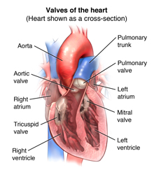
. There are two valves that are located between the atria and ventricles. There are four valves of the heart which are divided into two categories. Heart valves allow blood to go forward but not leak backwards.
These are bicuspid and tricuspid valves. The valve is composed of three leaflets working together to stop blood from entering the aorta prematurely. Prevents the backflow of blood as it is pumped from the left atrium to the left ventricle.
The aortic valve is the fourth and final heart valve lying between the left ventricle and the aorta. It then leaves the lungs to return to the heart through the pulmonary vein. The four valves of the heart allow blood into the heart and prevent it from flowing in the wrong direction.
The atria are the top two chambers of the heart that receive incoming blood from the body. These are similar to the strings supporting a parachute. They are located between the atria and.
The other two valves are located between the ventricles aorta and pulmonary arteries. Describe the sounds of each of the following heart valves in terms of intensity loudness pitch frequency and duration length. The valves keep the blood moving through the heart in the right direction.
The upper chambers the right and left atria receive incoming blood. - opens to allow blood to flow from the right atrium to the right ventricle. The heart has two types of valves that keep the blood flowing in the correct direction.
Part of the inside walls of the ventricles. Atrioventricular and semilunar valves. The valves between the atria and ventricles are called atrioventricular valves also called cuspid valves while those at the bases of the large vessels leaving the ventricles are called semilunar valves.
Separates the top right chamber right atrium from the bottom right chamber right ventricle. These four valves of our heart are. This is a subjective description.
It typically consists of three flaps or leaflets made of. For example tightness of the aortic valve is known as aortic stenosis. The lower chambers the more muscular right and left ventricles pump blood out of the heart.
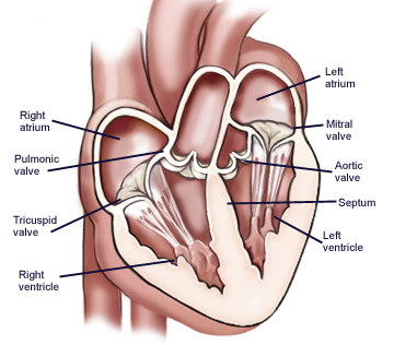
- opens to allow blood to pass from the left atrium to the left ventricle. A muscular wall known as the septum separates the two sides of the heart. Tightening of Heart Valves.
Between the right atrium and the right ventricle is the right atrioventricular valve or tricuspid valve. It is present on the opening of the post-caval vein inferior vena cava. When the valves become too tight it is known as stenosis.
They are composed of connective tissue and endocardium the inner layer of the heart. Opens to allow blood to flow from the right atrium to the right ventricle. The mitral valve The aortic valve The tricuspid valve The pulmonic valve.
The right atrium receives deoxygenated blood through the superior and inferior vena cavas from the body and pumps it to the right ventricle through the tricuspid valve which opens to allow the blood flow through and closes to prevent blood backing up the atrium. The valves open or close each. The Aortic Heart Valve.
These valves are flap-like tubes that prevent the backward flow of the blood. Page P-14 L17 Heart Sounds Biopac Student Lab 4 II. To prevent blood from flowing backwards back into the heart a system of one-way valves are present in the heart.
It drops through the mitral valve into the left ventricle. The bicuspid valve also known as the mitral valve is present. The four valves in order of circulation are.
The aortic valve opens when the ventricle contracts allowing blood to move from the heart and start the journey to the rest of the body. Prevents the backflow of blood as it is pumped from the right ventricle to the pulmonary artery. Begin with the aortic valve and compare others to it.
Has three leaflets or cusps. The left atrioventricular valve is the. Atrioventricular valves - The atrioventricular AV valves are located in the middle of the heart between the atria and ventricles and allow blood to flow from the atria into.
When the heart beats the ventricle pushes blood through the pulmonic valve into the pulmonary artery. The tricuspid valve and mitral bicuspid valve. View the full answer.
- located between the Right Artium and Right Ventricle. This controls the blood flow fr. Below are the four valves of the heart.
- located between the Left Artium and Left Ventricle. The heart valves which keep blood flowing in the right direction are gates at the chamber openings. A transverse section through the heart slightly above the level of the atrioventricular septum reveals all four heart valves along the same plane Figure 11.
The valves ensure unidirectional blood flow through the heart. The right atrioventricular valve is the tricuspid valve. Myocardium- This layer forms the heart muscles.
Right AV Valve aka Tricuspid Valve is between right atria and right ventricle Left AV Valve aka Bicuspid Valve aka Mitral Valve is between left atria and ventricle Chordae Tendonae - connect AV valves to the papillary muscles to keep valves from being floppy Semilunar Valves prevent backflow from the ventricles to the great arteries. Aortic Pulmonic Tricuspid Mitral. The heart valves can be broken down to two types.
The wall of the heart is made up of three layers. The heart have 4 valves which includes the mitral valve and tricuspid valve collectively called AV valve. Prevents the back flow of blood from.
The pulmonary artery carries blood to the lungs where it picks up oxygen. Stenosis is usually classified as mild moderate or severe. Prevents the backflow of blood as it is pumped from the left ventricle to the aorta.
It guards the opening of the coronary. Regurgitation can affect all 4 valves of the heart. Epicardium- This is a protective layer made of connective tissues.
These valves act as one-way inlets for blood located on the side of a chamber and one-way outlets for blood on the other side of a chamber. The valves of the heart are structures which ensure blood flows in only one direction. The human heart consists of mainly four heart valves.
These valves are tricuspid valve loca View the full answer.
4 Valves Of The Heart What Are They How They Work
Anatomy And Function Of The Heart Valves

Pumping Organ The Heart Heart Valves Anatomy Heart Organ Human Heart Function


0 Comments